File:DTI-sagittal-fibers.jpg

From Knowino
Jump to: navigation, search
Full resolution(1,021 × 1,125 pixels, file size: 203 KB, MIME type: image/jpeg)

This file is from a shared repository and may be used by other projects. The description on its file description page there is shown below.

Wikipedia

Cscr-featured.svgSound-icon-empty.svgThis is a featured picture on the German language Wikipedia (Exzellente Bilder) and is considered one of the finest images.
Cscr-featured.svgSound-icon-empty.svgThis is a featured picture on the English language Wikipedia (Featured pictures) and is considered one of the finest images.

If you think this file should be featured on Wikimedia Commons as well, feel free to nominate it.
If you have an image of similar quality that can be published under a suitable copyright license, be sure to upload it, tag it, and nominate it.


العربية | Azərbaycanca | Беларуская (тарашкевіца)‎ | বাংলা | Català | Čeština | Cymraeg | Dansk | Deutsch | Zazaki | Ελληνικά | English | Esperanto | Español | Eesti | فارسی | Suomi | Français | Galego | עברית | हिन्दी | Hrvatski | Magyar | Հայերեն | Bahasa Indonesia | Italiano | 日本語 | ქართული | Қазақша | 한국어 | Lëtzebuergesch | Lietuvių | Македонски | മലയാളം | Bahasa Melayu | Malti | Plattdüütsch | Nederlands | Norsk nynorsk | Norsk bokmål | Polski | Português | Português do Brasil | Română | Русский | Slovenščina | Српски / srpski | Svenska | தமிழ் | ไทย | Türkçe | Татарча/tatarça | Українська | Tiếng Việt | Yorùbá | 中文(简体)‎ | 中文(繁體)‎ | +/−

Summary[edit]

Description
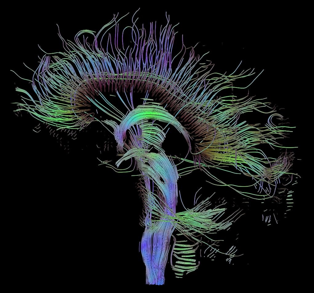
English: Visualization of a DTI measurement of a human brain. Depicted are reconstructed fiber tracts that run through the mid-sagittal plane. Especially prominent are the U-shaped fibers that connect the two hemispheres through the corpus callosum (the fibers come out of the image plane and consequently bend towards the top) and the fiber tracts that descend toward the spine (blue, within the image plane)
Français : Visualisation d'une mesure DTI d'un cerveau humain. Ce qui est représenté sont des faisceaux de fibres reconstruits qui traversent le plan demi-sagittal. On observe les fibres en U qui connectent les deux hémisphères à travers le corps calleux, qui sont particulièrement importantes (les fibres sortent du plan de l'image et par conséquent se courber vers le haut) ainsi que les faisceaux de fibres qui descendent vers la colonne vertébrale (bleu, dans le plan de l'image)
Deutsch: Traktographie-Verfahren rekonstruieren aus den Messdaten der Diffusions-Tensor-Bildgebung den anzunehmenden Verlauf größerer Nervenbahnen. Hier dargestellt sind die Ergebnisse für ein menschliches Gehirn; um die Übersichtlichkeit zu wahren, beschränkt sich die Abbildung auf Bahnen, die die Medianebene schneiden. Insbesondere sind dies die U-förmigen Faserbündel, die die beiden Hirnhälften verbinden (sie durchstoßen die Bildebene und sind nach oben gebogen) sowie die Faserbündel, die zum Rückenmark ziehen (blau dargestellt, liegen innerhalb der Bildebene)
Date
Source Own work
Author Thomas Schultz
Permission
(Reusing this file)

Rendering is own work, using a modified version of the BioTensor application developed at the University of Utah. The dataset is courtesy of Gordon Kindlmann at the Scientific Computing and Imaging Institute, University of Utah, and Andrew Alexander, W.M. Keck Laboratory for Functional Brain Imaging and Behaviour, University of Wisconsin, Madison. It is publicly available from [1]

Licensing[edit]

I, the copyright holder of this work, hereby publish it under the following licenses:
GNU head Permission is granted to copy, distribute and/or modify this document under the terms of the GNU Free Documentation License, Version 1.2 or any later version published by the Free Software Foundation; with no Invariant Sections, no Front-Cover Texts, and no Back-Cover Texts. A copy of the license is included in the section entitled GNU Free Documentation License.

w:en:Creative Commons
attribution share alike
This file is licensed under the Creative Commons Attribution-Share Alike 3.0 Unported license.
You are free:
  • to share – to copy, distribute and transmit the work
  • to remix – to adapt the work
Under the following conditions:
  • attribution – You must attribute the work in the manner specified by the author or licensor (but not in any way that suggests that they endorse you or your use of the work).
  • share alike – If you alter, transform, or build upon this work, you may distribute the resulting work only under the same or similar license to this one.
This licensing tag was added to this file as part of the GFDL licensing update.

w:en:Creative Commons
attribution share alike
This file is licensed under the Creative Commons Attribution-Share Alike 2.5 Generic, 2.0 Generic and 1.0 Generic license.
You are free:
  • to share – to copy, distribute and transmit the work
  • to remix – to adapt the work
Under the following conditions:
  • attribution – You must attribute the work in the manner specified by the author or licensor (but not in any way that suggests that they endorse you or your use of the work).
  • share alike – If you alter, transform, or build upon this work, you may distribute the resulting work only under the same or similar license to this one.

You may select the license of your choice.

File history

Click on a date/time to view the file as it appeared at that time.

Date/TimeThumbnailDimensionsUserComment
current16:22, 22 September 2006Thumbnail for version as of 16:22, 22 September 20061,021×1,125 (203 KB)Thomas Schultz ({{Information |Description=Visualization of a DTI measurement of a human brain. Depicted are reconstructed fiber tracts that run through the mid-sagittal plane. Especially prominent are the U-shaped fibers that connect the two hemispheres through the corp)

The following page links to this file:

Personal tools
Namespaces
Variants
Views
Actions
Navigation
Community
Toolbox