File:Magnetic resonance microscopy montage embryo.png
From Knowino
Size of this preview: 666 × 599 pixels
Full resolution (1,000 × 900 pixels, file size: 1.65 MB, MIME type: image/png)
Summary
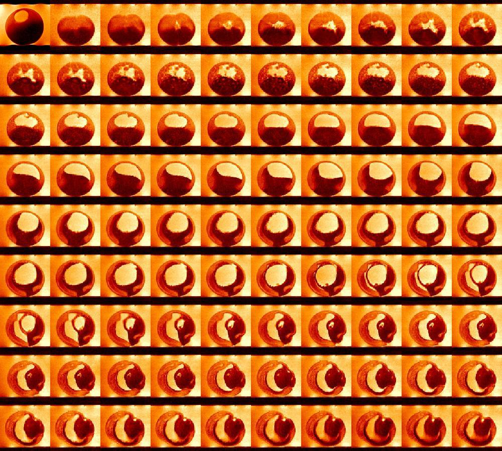
A montage of in vivo images acquired by means of magnetic resonance microscopy from a stage VI (prophase I-arrested) oocyte (top left frame) and the embryogenesis in the frog Xenopus laevis, from shortly after the first cell division until shortly prior to the hatching of the tadpole.
The top left frame was published in Fig. 1 of Lee et al., 2006. The remaining frames are from Movie 3 of Lee et al., 2007
Licensing
| |
This work is available under the terms of the Creative Commons Attribution–ShareAlike license. |
File history
Click on a date/time to view the file as it appeared at that time.
| Date/Time | Thumbnail | Dimensions | User | Comment | |
|---|---|---|---|---|---|
| current | 08:04, 8 August 2011 | | 1,000×900 (1.65 MB) | Paul Wormer (talk | contributions) | (A montage of in vivo images acquired by means of magnetic resonance microscopy from a stage VI (prophase I-arrested) oocyte (top left frame) and the embryogenesis in the frog Xenopus laevis, from shortly after the first cell division until shortly prior t) |
- Edit this file using an external application (See the setup instructions for more information)
File links
The following page links to this file: